

Enter this interactive experience to learn more about cytometry methods, how they work, and their capabilities and limitations.
Scroll to begin exploring Begin exploring


High-throughput single cell analysis helps uncover the layers of immune system complexity. New technologies offer the resolution to see the details, and the scale to see the big picture for researching the immune response.
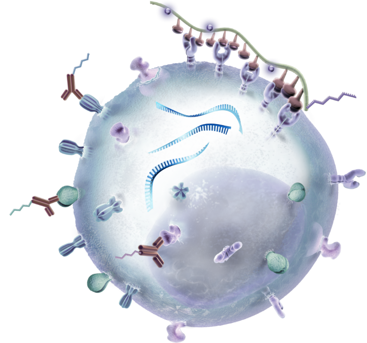
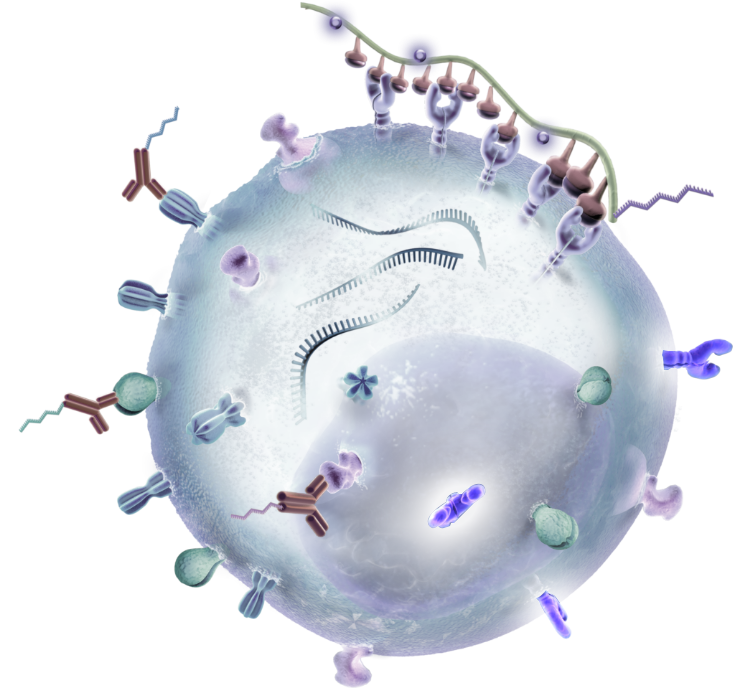
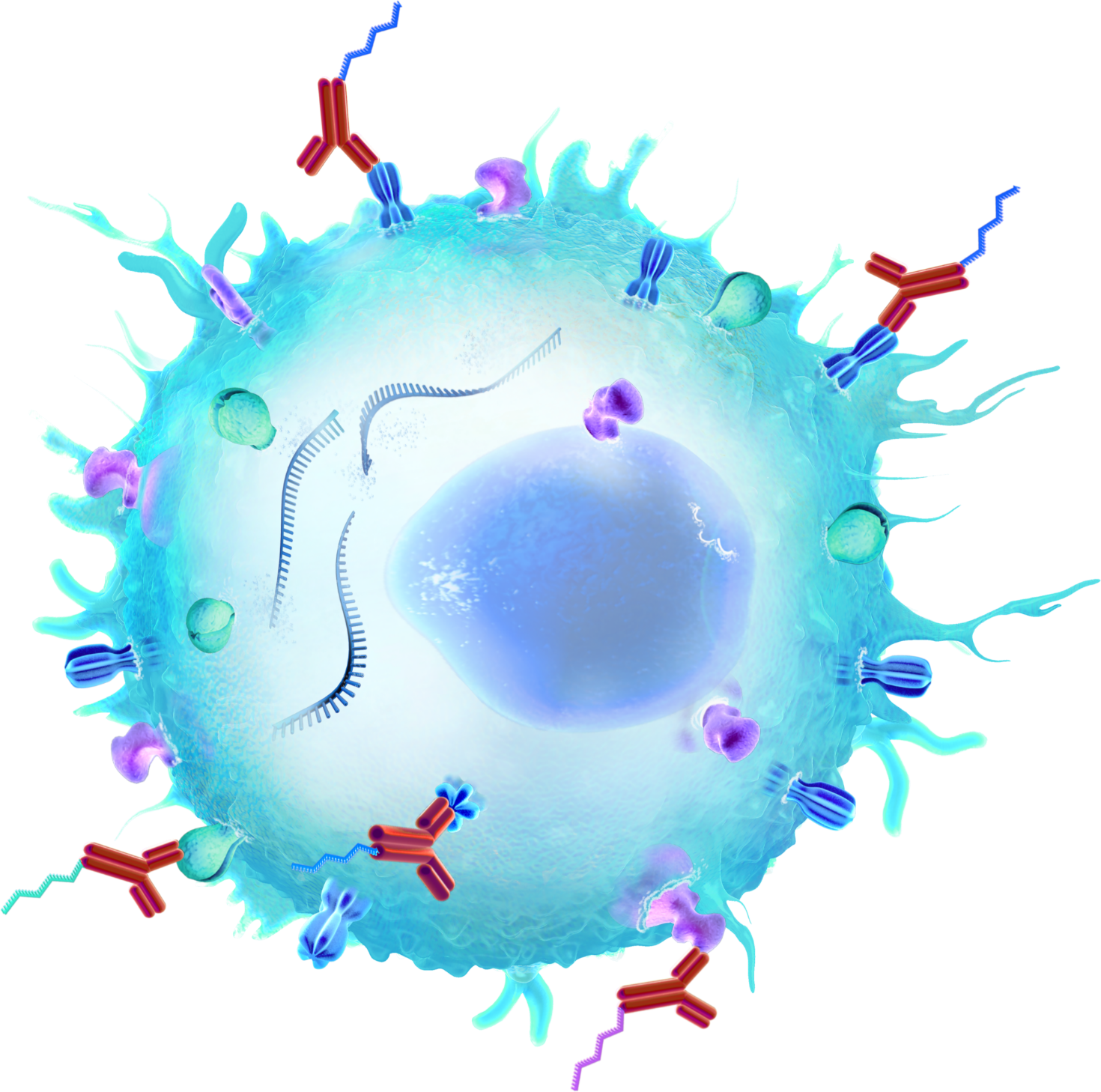
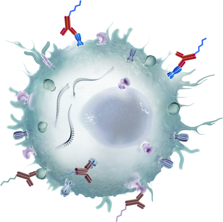
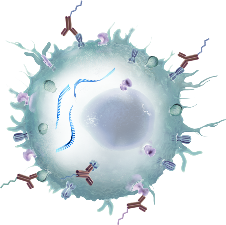
Specific cell markers are tagged with DNA barcodes, followed by single cell partitioning and barcoding, and next-generation sequencing.
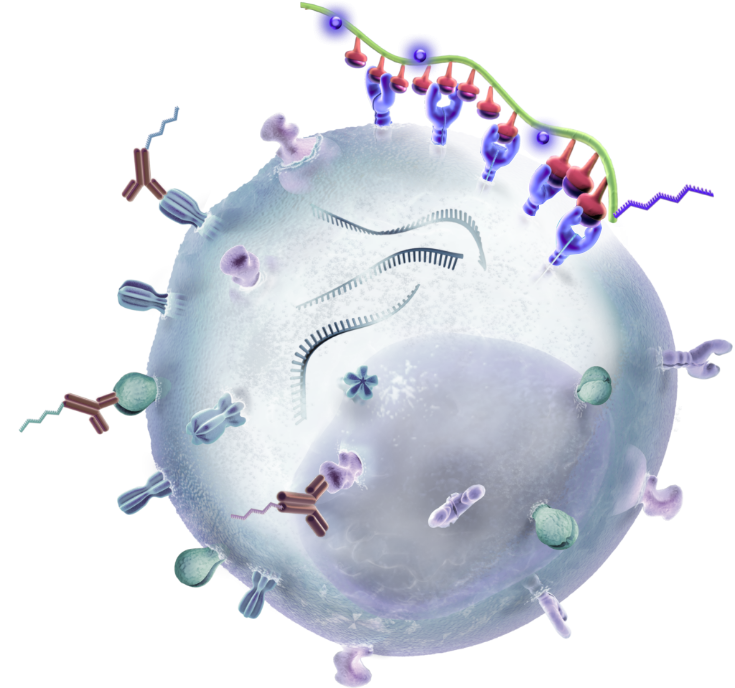
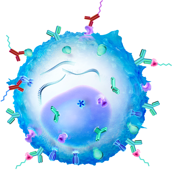
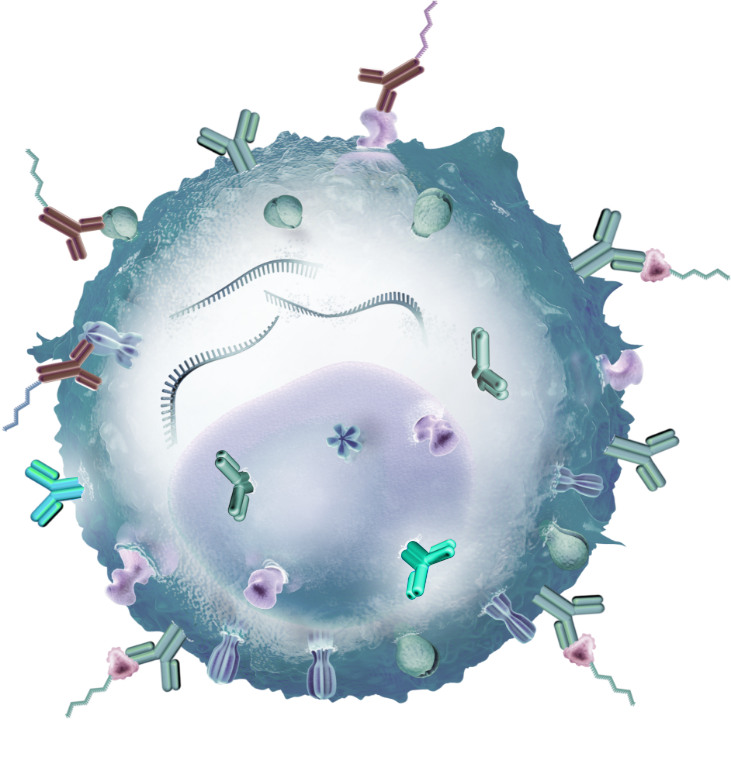
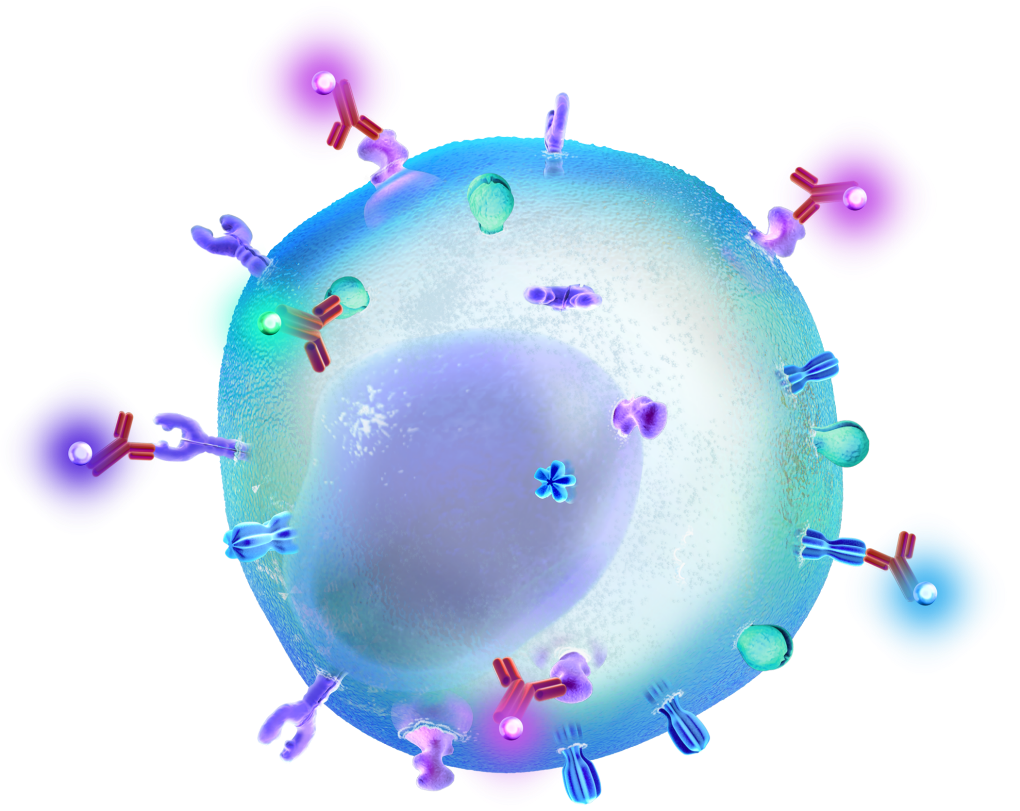
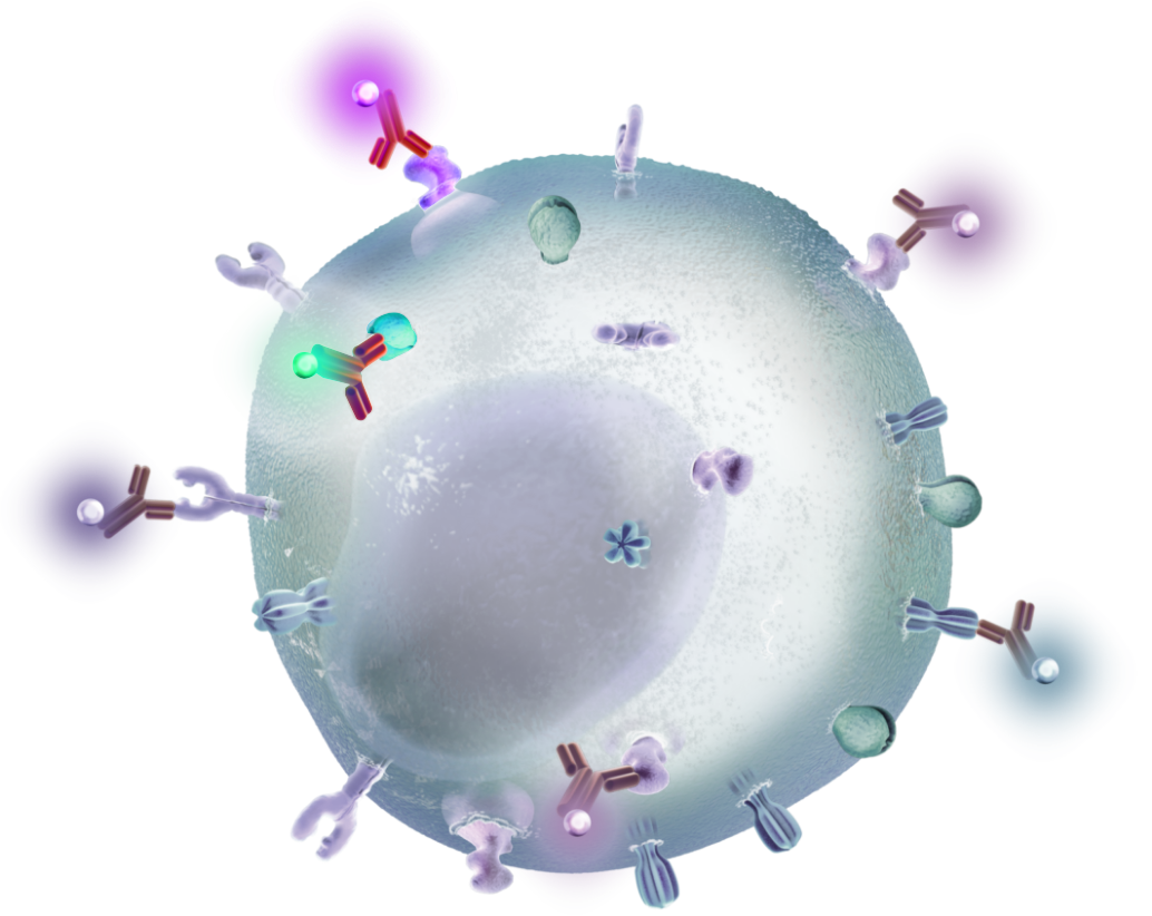
How Multiomic Cytometry worksSpecific cell markers are tagged with fluorescent dyes, then cells are counted or sorted as they flow one-at-a-time in a fluid stream through a beam of light.
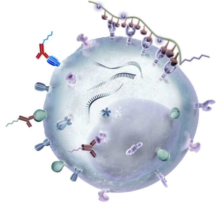
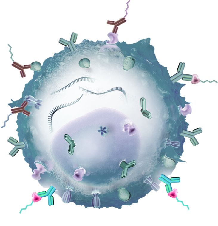
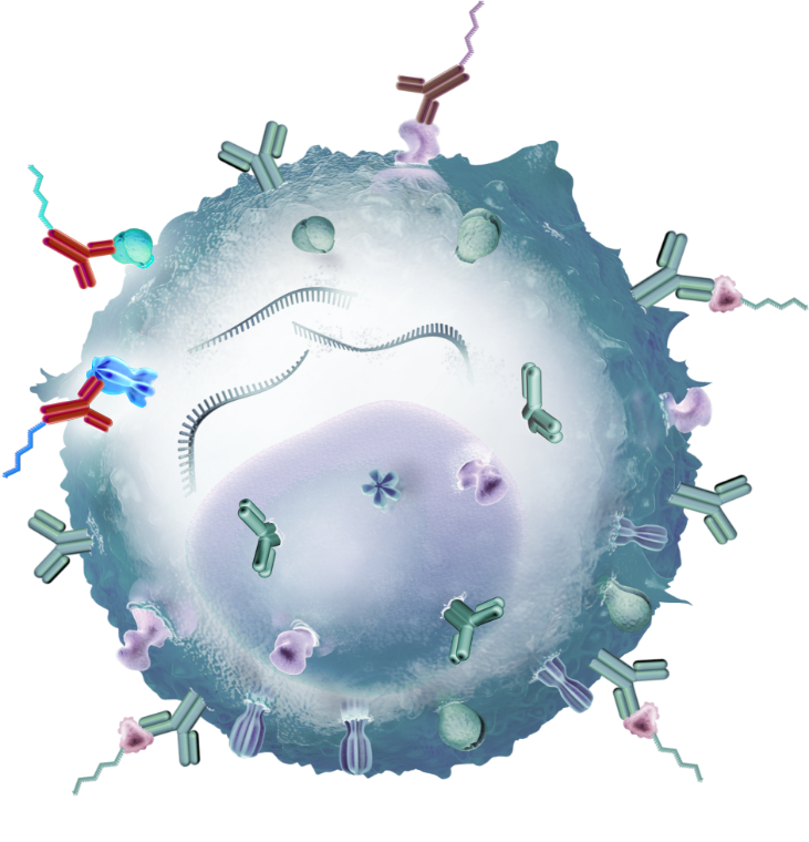
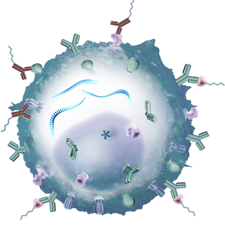
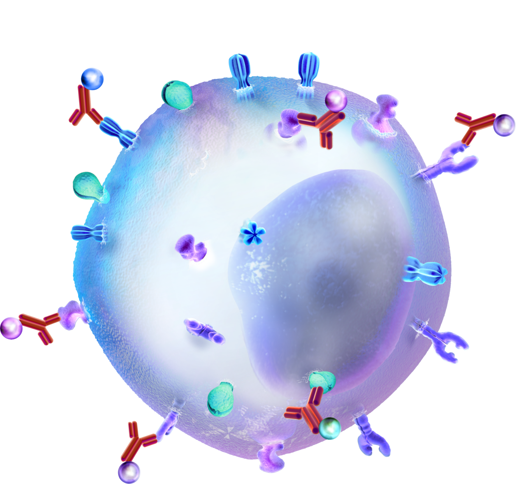
How Flow Cytometry worksSpecific cell markers are tagged with heavy metal isotopes, then cells are vaporized and metal ions are counted on a time-of-flight (TOF) mass spectrometer.
How Mass Cytometry works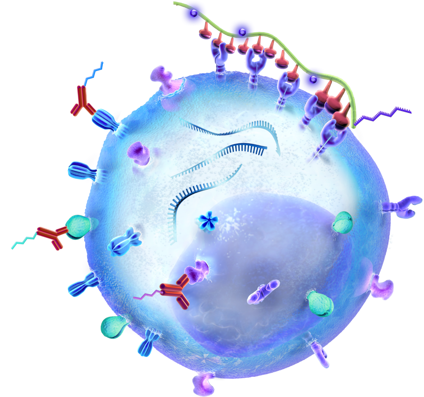
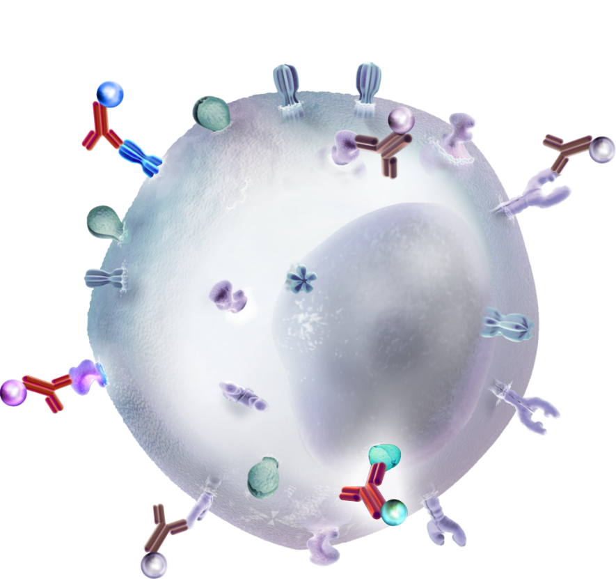
High-throughput single cell analysis helps uncover the layers of immune system complexity. New technologies offer the resolution to see the details, and the scale to see the big picture for researching the immune response.
The immune system is a complex network of cell types, all working together to maintain health and eliminate pathogens. Characterizing the dynamics of the immune response requires the ability to phenotype individual cells at massive scale, using multiple parameters. To this end, sophisticated methods for high-throughput single cell analysis provide for increasingly fine-tuned immune profiling.
Technologies like flow cytometry, mass cytometry, and multiomic cytometry use cell surface proteins as markers to help distinguish immune cell subtypes and activation states. Additional technologies are expanding the immunolgist’s toolbox for identifying new biomarkers, designing immunotherapies, and monitoring the antigen-specific response.
Watch this short animation to learn how you can explore immunology with unprecedented breadth, depth, and precision.

Multiomic cytometry can measure cell surface epitopes, mRNA transcripts, antigen receptors and their cognate antigens, and more, all at the same time. Multiomic cytometry makes use of DNA barcodes, conjugated to other biomolecules, to measure the existence and prevalence of specific biomolecular analytes by turning a next-generation sequencer into an ultra-high parameter cytometric detector.
DNA barcode labels overcome the limitations of traditional cytometric methods
For example, DNA barcodes conjugated to antibody reagents can enable the simultaneous labeling and analysis of up to hundreds of cell surface proteins, removing the constraint on simultaneous epitope analysis imposed by the spectral overlaps of fluors or metal ions. DNA barcodes can similarly be used to label large panels of antigens or peptide-MHC molecules to screen for antigen specificity in B or T cells. Many other applications of this technology can, of course, be imagined and designed for other cellular features and diverse additional differentiated cell types.
True high parameter analysis provides full characterization of the immune system
Multiomic cytometry via single cell sequencing allows immunologists to simultaneously assay hundreds to thousands of cell-surface and intracellular mRNA-based gene expression parameters in hundreds to tens-of-thousands of individual cells. This information can be matched with the paired, full-length V(D)J sequences of each antigen receptor clonotype. Single cell transcriptomes from 10x Genomics can be based on unbiased whole transcriptomes or targeted gene expression panels of interest. Bioinformatics software maps each data point back to each single cell. This comprehensive cellular phenotyping provides the scale and resolution to fully characterize the diverse cell states and dynamics of the immune system.
In many cases, researchers may consider using flow cytometry first to enrich for specific cell types.
Watch this webinar to learn about how to apply multiomic cytometry to infectious disease research Download our comprehensive Immunology Research Guide and learn more about applying single cell sequencing to address the biggest topics in immunology


Single cell suspensions are generated and stained with antibodies (or other ligands) that are tagged with different color fluorescent dyes. In the flow cytometer, cells flow single file, thousands-per-second past a laser. The light scatter and the emitted fluorescence recorded for each cell can determine characteristics like cell size, complexity, and protein expression. To distinguish clear signals, computers mathematically compensate for spectral overlap and autofluorescence from cells. Although many fluorophore colors exist, it is difficult to do compensation for more than 8 colors at once. Cells can be counted, or sorted and recovered for downstream analysis.
Are you looking for better ways to perform multi-parametric analysis? Learn more about how to measure cell surface proteins, antigen specificity and gene expression by downloading this application note.
Learn more
Cells are fixed and stained with a panel of antibodies conjugated to stable heavy metal isotopes. In the mass cytometer, single cell droplets travel through a plasma stream, where cells get vaporized into a cloud of ions. The ion cloud is filtered and the TOF mass spectrometer separates the metal isotopes by their precise mass-to-charge ratio. The per-cell number of each metal tag indicates levels of protein expression. This method can also measure intracellular signalling pathways using phospho-specific antibodies. Due to the breadth of stable heavy metals of different atomic weights, mass cytometry allows analysis of over 40 cellular features at once.
Are you looking for better ways to perform multi-parametric analysis? Learn more about how to measure cell surface proteins, antigen specificity, and gene expression simultaneously by watching this webinar.
Learn more
The adaptive immune response is controlled, in part, by major histocompatibility (MHC) cell surface proteins presenting antigens to T cells. In B cells, the antigen-recognizing immunoglobulins (Ig) exist in either membrane-bound or secreted forms, known as B cell receptors (BCRs) or antibodies (Ab), respectively.
Leverage the power of integrating next-generation sequencing to measure antigen specificity
Single cell sequencing can include panels of peptide-MHC multimers tagged with DNA barcodes to profile the antigen specificity of hundreds to tens-of-thousands of T lymphocytes in a single experiment. Similarly, antigens tagged with DNA barcodes can be used to confirm the antigen specificity of BCRs. This same single cell sequencing analysis can simultaneously measure transcriptomes, cell surface epitopes, and detailed immune V(D)J repertoires, providing the most comprehensive means of assessing adaptive immune clonality, specificity, and phenotype in a single experimental measurement.
Using flow cytometry to sort samples first and enrich for immune cells of interest can increase the potential to detect rare cell types.
Explore the single cell immune profiling workflow and multiomic data generated in a study of PBMCs from two donors infected by cytomegalovirus by watching this webinar.
Watch how it works: See how multiomic cytometry allows you to map immunity in greater detail on a cell by cell basis.


Single cell sequencing leverages antibodies conjugated to DNA barcodes, as an alternative to the fluorescent tags used in flow cytometry. Barcode diversity vastly expands the number of cell surface markers that can be analyzed, and offers highly multiplexed information for hundreds to tens-of-thousands of cells. Researchers may use flow cytometry to sort samples first and enrich for cells of interest. This focuses sequencing resources and expands the ability to detect rare cell types.
Deeply characterize cell types by combining cell surface protein and RNA expression
Single cell sequencing combines cell surface protein detection with an unbiased readout of each single cell transcriptome. Layering protein expression data over digital gene expression data, on a cell-by-cell basis, offers a comprehensive view of cell states. Additional parameters, such as clonotype and antigen specificity, easily integrate into the same single cell sequencing assay.
Watch how it works: See how multiomic cytometry allows you to map immunity in greater detail on a cell by cell basis.

Transcription, gene expression, and molecular regulatory mechanisms are biological features of single cells. Deepening our understanding of biology necessarily requires that we study biological processes at this fundamental level.
Single cell RNA sequencing for unbiased characterization of cell populations
While the number of transcripts sequenced per sample are similar between single cell RNA-seq and bulk expression, bulk analysis averages this gene expression measurement across the number of cells that were present in the sample, prior to analysis. Single cell gene expression studies, therefore, allow you to extend beyond traditional global marker gene analysis to the characterization of cell types or cell states and the concomitant dynamic changes in regulatory pathways, which are driven by many genes at a cellular level. Importantly, single cell gene expression allows for an unbiased characterization of cell populations independently of any prior knowledge of cell subtypes or cell markers.
Identify rare cell types and define new cell sub-types
Single cell transcriptomic technologies allow the direct measurement of gene expression at the single cell level to quantify inter-cellular population heterogeneity and characterize cell types, cell states, and dynamic cellular transitions cell by cell. In addition to potentially identifying new cell subtypes and rare cell populations, single cell technologies enable a better understanding of transcription dynamics and gene regulatory relationships.
Whether you have questions about how to design your experiments, optimize parameters, or identify the computational/analytical tools necessary to best analyze your single cell gene expression data, this guide has you covered. Take full advantage of the rich information enabled by single cell transcriptomic technology by downloading the getting started guide today.
Learn more
The specificity of adaptive immunity relies on the vast array of unique lymphocyte receptors generated through recombination of Variable (V), Diversity (D), and Joining (J) gene segments. Single cell immune profiling captures paired, full-length V(D)J sequences for T-cell receptor and B-cell immunoglobulin genes for hundreds to tens-of-thousands of individual cells.
Multiomic phenotyping combined with immune profiling for enhanced cell phenotyping
This unbiased analysis can characterize clonotype diversity on a cell-by-cell basis. Cell sorting with flow cytometry, followed by single cell immune profiling, can increase the power to detect rare clonotypes by enriching samples for adaptive immune cell populations to more deeply profile them.
Multiomic cytometry via single cell sequencing can assay multiple dimensions of immune cell function with high resolution and scale. Immunologists can now study the clonality and diversity of the adaptive immune system in any sample, and pair these measurements with the most detailed multiomic single-cell cytometric phenotype of the same cells.
Profile the Adaptive Immune System
Watch how it works: Single cell immune sequencing
unlocks multiple dimensions of immune cell function.
Learn more about how to identify cell type-specific
immune repertoires on a cell-by-cell basis.

